

Introduction
Understanding dog lung anatomy is essential for anyone working in veterinary practice. The respiratory system is one of the most frequently assessed body systems in clinical cases, from coughing dogs in consults to emergency respiratory distress.
For veterinary nurses and students, a solid grasp of how the lungs are structured and how they function is key to accurate monitoring, effective nursing care, and supporting diagnostic procedures.
This guide breaks down dog lung anatomy in a clear, practical way that links structure to clinical relevance.
Read also: Heart Anatomy and Physiology for Veterinary Nurses: An Essential Clinical Guide – Paws and Pet Hub
Overview of Dog Lung Anatomy
The lungs are a pair of spongy organs located within the thoracic cavity, protected by the rib cage. Their main role is gas exchange—bringing oxygen into the body and removing carbon dioxide.
In dog lung anatomy, each lung is divided into lobes, with a branching airway system that ensures air reaches deep into the alveoli where gas exchange occurs.
The lungs work closely with:
- Trachea
- Bronchi and bronchioles
- Diaphragm
- Rib muscles
Together, these structures allow efficient breathing and oxygen delivery.
The Airway Pathway in Dogs
Air enters the respiratory system in a highly structured sequence:
- Nasal cavity or mouth
- Pharynx
- Larynx
- Trachea
- Primary bronchi
- Smaller bronchi and bronchioles
- Alveoli
Each step in this pathway is designed to filter, warm, and humidify air before it reaches the delicate lung tissue.
From a nursing perspective, understanding this pathway helps localise disease—for example, upper airway noise vs lower airway crackles.
Lung Lobes in Dog Lung Anatomy
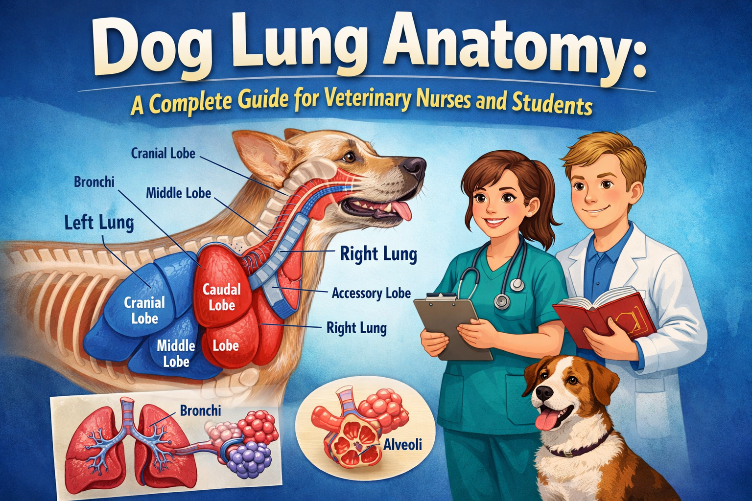
One of the most important features of dog lung anatomy is the division into lobes.
Right lung lobes:
- Cranial lobe
- Middle lobe
- Caudal lobe
- Accessory lobe
Left lung lobes:
- Cranial lobe (divided into cranial and caudal parts)
- Caudal lobe
This asymmetry is clinically important during imaging and surgery. For example, aspiration pneumonia often affects the right cranial and middle lobes due to airway anatomy and gravity.
Alveoli: Where Gas Exchange Happens
The alveoli are tiny air sacs at the end of the respiratory tree. They are the functional units of the lungs.
In dog lung anatomy, alveoli are surrounded by capillaries, allowing oxygen and carbon dioxide to diffuse across thin membranes.
Key features:
- Extremely large surface area
- Thin epithelial walls
- Rich blood supply
Damage to alveoli (e.g. pneumonia or oedema) significantly impairs breathing efficiency.
Pleura and Thoracic Protection
The lungs are enclosed by a double-layered membrane called the pleura:
- Visceral pleura (covers the lungs)
- Parietal pleura (lines the chest wall)
A small amount of pleural fluid allows smooth movement during breathing.
Conditions affecting the pleural space, such as pneumothorax or pleural effusion, can severely restrict lung expansion and are often emergencies in practice.
How Dog Lungs Work (Physiology Overview)
Breathing involves two main processes:
Inhalation
- Diaphragm contracts and flattens
- Rib muscles expand the chest cavity
- Pressure decreases, drawing air into the lungs
Exhalation
- Diaphragm relaxes
- Chest cavity volume decreases
- Air is expelled from the lungs
Although breathing isn’t purely a physics topic, the movement of air is driven by pressure gradients created by muscular and thoracic changes.
From a clinical nursing perspective, any pain, fluid accumulation, or airway obstruction can disrupt this balance.
Clinical Relevance for Veterinary Nurses
Understanding dog lung anatomy is not just theoretical—it directly supports clinical practice.
1. Respiratory assessment
- Listening for crackles, wheezes, or silence
- Identifying abnormal breathing patterns
2. Oxygen therapy
Knowledge of lung structure helps understand why oxygen delivery is essential in hypoxic patients.
3. Imaging interpretation
Radiographs depend on recognising lung lobes and changes in lung opacity.
4. Emergency care
Conditions like:
- Pulmonary oedema
- Pneumonia
- Pneumothorax
require rapid anatomical understanding for effective intervention.
Common Conditions Affecting Dog Lungs
Pneumonia
Inflammation or infection of lung tissue, often affecting dependent lobes.
Pulmonary oedema
Fluid accumulation in alveoli, commonly linked to heart disease.
Bronchitis
Inflammation of airways leading to chronic coughing.
Collapsed lung (atelectasis)
Partial or complete collapse of lung tissue due to obstruction or compression.
Each of these conditions alters normal dog lung anatomy function, affecting oxygen exchange.
Why Dog Lung Anatomy Matters in Veterinary Nursing
For veterinary nurses, understanding this anatomy improves:
- Patient monitoring accuracy
- Early detection of deterioration
- Interpretation of clinical signs
- Support during diagnostic imaging
- Emergency response confidence
It also strengthens communication with vets during case discussions and treatment planning.
Frequently Asked Questions
How many lung lobes do dogs have?
Dogs typically have five main lung lobes, with the left cranial lobe divided into two parts.
Where does gas exchange occur in dog lungs?
Gas exchange occurs in the alveoli.
Why is dog lung anatomy important in nursing?
It helps with assessment, monitoring, emergency care, and understanding respiratory disease.
Final Thoughts
A strong understanding of dog lung anatomy is essential for veterinary nurses and students working in small animal practice. It underpins everything from routine respiratory checks to critical care decision-making.
When you understand how the lungs are structured and how they function, you can better recognise when something goes wrong—and respond faster when it matters most.
Read also: The Dog Respiratory System Anatomy – Lungs and Respiratory Tract



Leave a Reply