

Introduction
Understanding kidney anatomy in dogs is essential for veterinary nurses, particularly when managing patients with renal disease, urinary issues, dehydration, or systemic illness. The kidneys play a vital role in maintaining homeostasis, filtering waste products from the blood, regulating electrolytes, and controlling fluid balance.
In veterinary practice, renal dysfunction is a common clinical presentation, especially in older dogs. A strong understanding of canine kidney structure and function helps veterinary nurses monitor patients more effectively, assist in diagnostics, and provide appropriate nursing care.
This guide breaks down the anatomy of the canine kidney in a clear, practical way designed specifically for veterinary professionals.
Also read: Heart Anatomy and Physiology for Veterinary Nurses: An Essential Clinical Guide
Overview of the Canine Urinary System
The urinary system consists of:
- Kidneys
- Ureters
- Urinary bladder
- Urethra
The kidneys are the primary functional organs responsible for urine production. They filter blood, remove metabolic waste, and regulate water, electrolytes, and acid-base balance.
In dogs, the kidneys are bean-shaped organs located in the retroperitoneal space, meaning they sit behind the abdominal cavity.
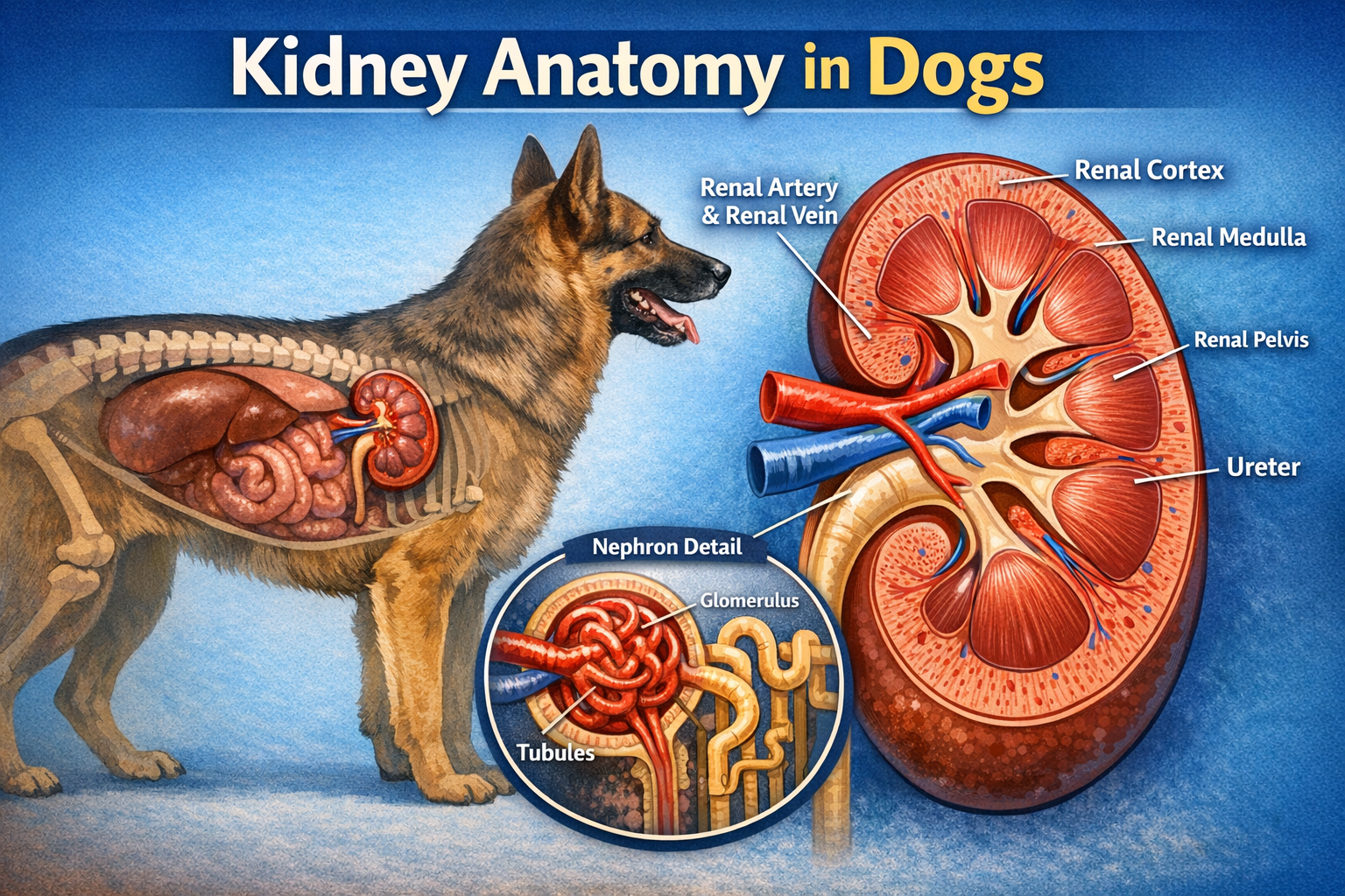
Kidney Anatomy in Dogs
The canine kidney structure can be divided into several key components:
1. Renal Capsule
A thin, fibrous protective layer that surrounds the kidney. It helps maintain shape and protects internal structures from trauma and infection.
2. Cortex
The outer region of the kidney. The cortex contains:
- Glomeruli
- Proximal and distal convoluted tubules
This is where initial blood filtration begins.
3. Medulla
The inner region of the kidney, made up of:
- Renal pyramids
- Loop of Henle structures
- Collecting ducts
The medulla plays a major role in concentrating urine.
4. Renal Pelvis
A funnel-shaped structure that collects urine from the collecting ducts and channels it into the ureter.
5. Hilus
The entry and exit point for:
- Renal artery
- Renal vein
- Ureter
Blood Supply of the Kidneys: Kidney Anatomy in Dogs
The kidneys receive a high volume of blood flow, reflecting their role in filtration.
Key vessels include:
- Renal artery (from abdominal aorta)
- Segmental arteries
- Interlobar arteries
- Afferent arterioles → glomeruli
- Efferent arterioles
- Peritubular capillaries and vasa recta
- Renal vein → caudal vena cava
This complex vascular system ensures efficient filtration and reabsorption.
💡 Clinical relevance: Any disruption in renal blood flow can rapidly lead to acute kidney injury.
The Nephron: Functional Unit of the Kidney
The nephron anatomy in dogs is crucial for understanding renal physiology.
Each kidney contains thousands of nephrons, responsible for filtering blood and producing urine.
Components of the Nephron:
1. Glomerulus
A network of capillaries where blood filtration begins. Small molecules pass into Bowman’s capsule.
2. Bowman’s Capsule
Surrounds the glomerulus and collects the filtrate.
3. Proximal Convoluted Tubule (PCT)
Reabsorbs:
- Water
- Glucose
- Amino acids
- Electrolytes
This is the major reabsorption site.
4. Loop of Henle
Responsible for concentrating urine. It creates a salt gradient in the medulla.
5. Distal Convoluted Tubule (DCT)
Fine-tunes electrolyte balance and pH regulation.
6. Collecting Duct
Final adjustment of urine concentration before it enters the renal pelvis.
Types of Nephrons in Dogs: Kidney Anatomy in Dogs
Dogs have two main types of nephrons:
Cortical Nephrons
- Located in the outer cortex
- Short loops of Henle
- Responsible for most filtration
Juxtamedullary Nephrons
- Located near the medulla
- Long loops of Henle
- Essential for urine concentration
Kidney Function in Dogs: Kidney Anatomy in Dogs
Understanding dog kidney function is essential in veterinary nursing.
The kidneys perform several vital roles:
1. Filtration of Blood
Removes waste products such as:
- Urea
- Creatinine
- Toxins
2. Fluid Balance
Regulates hydration status by adjusting urine output.
3. Electrolyte Regulation
Controls:
- Sodium
- Potassium
- Calcium
- Phosphorus
4. Acid-Base Balance
Maintains blood pH within a narrow range.
5. Hormone Production
The kidneys produce:
- Erythropoietin (stimulates red blood cell production)
- Renin (blood pressure regulation)
- Calcitriol (active vitamin D)
Clinical Relevance for Veterinary Nurses
A strong understanding of kidney anatomy in dogs is essential in practice.
Common Conditions:
- Chronic kidney disease (CKD)
- Acute kidney injury (AKI)
- Pyelonephritis
- Congenital renal dysplasia
Nursing Considerations:
- Monitoring urine output
- Fluid therapy management
- Blood pressure monitoring
- Biochemistry interpretation (urea, creatinine, SDMA)
- Monitoring hydration status
Key Warning Signs:
- Polyuria/polydipsia
- Weight loss
- Vomiting
- Lethargy
- Poor appetite
Why Kidney Anatomy in Dogs Matters in Veterinary Nursing
Veterinary nurses play a critical role in early detection and management of renal disease. Understanding the canine renal system explained simply allows for:
- Faster recognition of abnormalities
- Better patient monitoring
- Improved communication with vets
- More accurate nursing care plans
Kidney disease is often progressive, meaning early intervention can significantly improve outcomes.
Summary
The anatomy of the kidney in dogs is a complex but essential topic for veterinary nurses. From the cortex and medulla to the nephron’s filtration system, each structure plays a vital role in maintaining homeostasis.
A solid understanding of renal anatomy improves clinical confidence and enhances patient care, particularly in cases of chronic or acute kidney disease.
Useful Link: Canine Renal Ultrasound: A Guide to Interpretation | Royal Canin Academy





Leave a Reply