
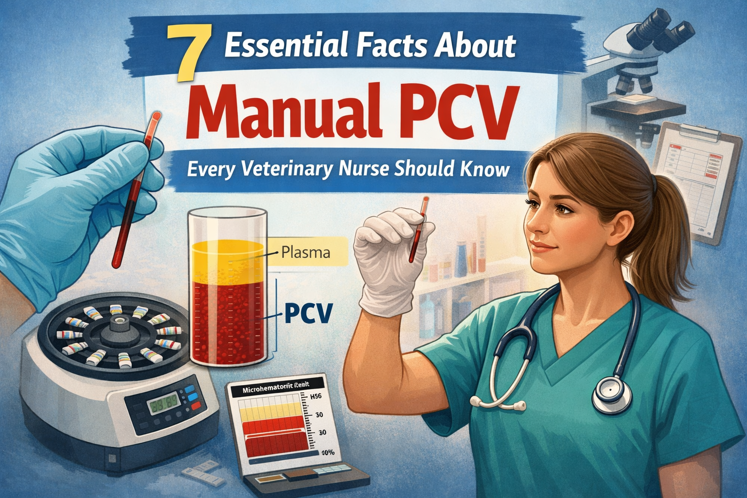

Packed cell volume (PCV) is a simple but incredibly valuable diagnostic test used every day in veterinary practice. Whether assessing an emergency patient, monitoring anaemia, or evaluating hydration status, understanding how to perform and interpret a manual PCV is an essential skill for veterinary nurses and veterinary professionals.
Although many practices use automated haematology analysers, manual PCV testing remains a reliable, quick, and cost-effective method that can provide important clinical information when time is critical.
In this guide, we’ll explore what is a PCV in veterinary medicine, how to perform a PCV test, what results mean, and why understanding PCV values is essential in patient care.
Read also: IV Catheter Placement in Dogs 10 Step Veterinary Nurse Guide
What Is a PCV in Veterinary Medicine?
So, what is a PCV in veterinary medicine?
PCV stands for packed cell volume and refers to the percentage of blood made up of red blood cells (erythrocytes).
When whole blood is centrifuged, the components separate into three layers:
- Packed red blood cells – the bottom layer
- Buffy coat – a thin white layer containing white blood cells and platelets
- Plasma – the clear fluid layer at the top
The PCV measures how much of the blood volume consists of red blood cells.
For example:
A PCV of 40% means that 40% of the blood volume is made up of red blood cells, while the remaining 60% consists mainly of plasma.
Why Is Manual PCV Important in Veterinary Practice?
A manual PCV veterinary test can provide rapid information about a patient’s red blood cell status.
It is commonly used for:
- Emergency patients
- Trauma cases
- Blood loss assessment
- Anaemia monitoring
- Fluid therapy assessment
- Pre-anaesthetic checks
- Critical care monitoring
A PCV is often performed alongside total protein (TP), giving clinicians additional information about hydration status and potential disease processes.
How to Perform a PCV Test in Veterinary Practice
Knowing how to perform a PCV test in veterinary settings is an important practical skill.
The basic steps include:
1. Collect a Blood Sample
A blood sample is collected into an appropriate anticoagulant tube, usually containing EDTA.
Ensure the sample is mixed gently to prevent clot formation.
2. Fill the Haematocrit Tube
Using a capillary tube:
- Fill approximately two-thirds to three-quarters of the tube with blood
- Avoid introducing air bubbles
- Seal one end using sealant or clay
3. Centrifuge the Sample
Place the tube into a microhaematocrit centrifuge.
The centrifuge separates the blood components, allowing the red blood cells to pack together.
4. Read the PCV Result
The percentage of packed red cells is measured using a PCV reader or haematocrit chart.
The reading is taken from:
- The bottom of the packed red cell layer
- To the top of the plasma layer
The buffy coat is not included in the PCV measurement.
What Is a Normal PCV in Animals?
A common question veterinary professionals ask is: what is a normal PCV in animals?
Reference ranges vary between species, breeds, age groups, and laboratories.
Approximate normal ranges include:
Dogs:
35–55%
Cats:
30–45%
Horses:
32–48%
Rabbits:
33–50%
Always interpret PCV alongside the patient’s clinical signs and laboratory findings.
What Is the Normal PCV for a Healthy Dog?
So, what is the normal PCV for a healthy dog?
A healthy adult dog will typically have a PCV around:
35–55%
However, breed variation exists. For example, some sighthounds may naturally have higher PCV values than other breeds.
A result outside the reference range does not automatically indicate disease — it should always be interpreted alongside history, examination findings, and other diagnostics.
Can Dehydration Increase PCV?
Yes, can dehydration increase PCV? Absolutely.
Dehydration reduces the amount of plasma within the bloodstream, causing the blood to become more concentrated.
This can result in a falsely elevated PCV, known as relative erythrocytosis.
A dehydrated patient may have:
- Increased PCV
- Increased total protein
- Reduced circulating fluid volume
This is why PCV should never be interpreted alone.
A patient with a high PCV may not necessarily have an increased number of red blood cells — they may simply have less plasma.
What Happens If the PCV Is High?
A high PCV indicates that there is an increased proportion of red blood cells within the blood.
Potential causes include:
Dehydration
The most common cause of increased PCV.
Stress response
Excitement or fear can cause temporary changes due to splenic contraction.
Chronic oxygen deficiency
Conditions causing reduced oxygen delivery may stimulate increased red blood cell production.
Primary or secondary erythrocytosis
Less common disorders can cause true increases in red blood cell numbers.
So, what happens if the PCV is high?
The clinical significance depends on the cause. A mildly increased PCV in a dehydrated patient may resolve with fluid therapy, while persistent increases require further investigation.
What Is a Dangerously Low PCV in Dogs?
A low PCV indicates anaemia, meaning the patient has fewer circulating red blood cells.
So, what is a dangerously low PCV in dogs?
While severity depends on the individual patient, a PCV below approximately:
20–25%
is generally considered clinically significant and may require further investigation or intervention.
A severely low PCV, particularly:
below 15–20%
may indicate life-threatening anaemia, especially if the patient is showing clinical signs.
Signs associated with anaemia may include:
- Pale gums
- Weakness
- Lethargy
- Increased heart rate
- Collapse
- Exercise intolerance
Causes of a Low PCV
A reduced PCV may occur due to:
Blood Loss
Examples include:
- Trauma
- Surgery
- Internal bleeding
- Parasites
Reduced Red Blood Cell Production
Causes may include:
- Bone marrow disease
- Chronic illness
- Nutritional deficiencies
Increased Red Blood Cell Destruction
Known as haemolysis, this can occur with:
- Immune-mediated haemolytic anaemia (IMHA)
- Toxins
- Infectious diseases
PCV and Total Protein: Why They Are Often Checked Together
PCV and total protein provide valuable information when interpreted together.
Examples:
High PCV + High Total Protein
Often suggests dehydration.
Low PCV + Low Total Protein
May indicate blood loss.
Low PCV + Normal/High Total Protein
May suggest processes such as inflammation or immune-mediated disease.
These values help veterinary teams build a clearer picture of what is happening within the patient.
Common Errors When Performing Manual PCV Testing
Even though PCV testing is straightforward, mistakes can affect accuracy.
Common issues include:
Incorrect sample mixing
Clotted samples may produce inaccurate results.
Underfilling tubes
Insufficient blood volume can affect reliability.
Poor centrifugation
Incomplete separation may result in incorrect readings.
Including the buffy coat
The buffy coat should not be included when measuring PCV.
Final Thoughts
A manual PCV remains an essential diagnostic skill for veterinary nurses and clinical teams. Despite advances in automated analysers, the ability to quickly perform and interpret a PCV test can provide valuable information during emergencies and routine patient care.
Understanding what is a PCV in veterinary medicine, knowing how to perform a PCV test in veterinary practice, and recognising abnormal results allows veterinary professionals to make informed decisions and provide better patient support.
Whether monitoring anaemia, assessing hydration, or evaluating a critical patient, manual PCV testing remains a simple technique with significant clinical value.
Read also: PCV University of Bristol




Leave a Reply